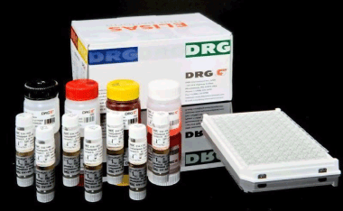
Estradiol ELISA Kits
Biozatix News – Informasi Popular Sains Teknologi dan Kesehatan

Estradiol (1,3,5(10)-estratriene-3,17β-diol; 17β-estradiol; E2) is a C18 steroid hormone with a phenolic A ring. This steroid hormone has a molecular weight of 272.4. It is the most potent natural Estrogen, produced mainly by the Graffian follicle of the female ovary and the placenta, and in smaller amounts by the adrenals, and the male testes (1,2,3). Estradiol (E2) is secreted into the blood stream where 98% of it circulates bound to sex hormone binding globulin (SHBG) and to a lesser extent to other serum proteins such as albumin. Only a small fraction circulates as free hormone or in the conjugated form (4,5). Estrogenic activity is affected via estradiol-receptor complexes which trigger the appropriate response at the nuclear level in the target sites. These sites include the follicles, uterus, breast, vagine, urethra, hypothalamus, pituitary and to a lesser extent the liver and skin. In non-pregnant women with normal menstrual cycles, estradiol secretion follows a cyclic, biphasic pattern with the highest concentration found immediately prior to ovulation (6,7). The rising estradiol concentration is understood to exert a positive feedback influence at the level of the pituitary where it influences the secretion of the gonadotropins, follicle stimulating hormone (FSH), and luteinising hormone (LH), which are essential for folicular maturation and ovulation, respectively (8,9). Following ovulation, estradiol levels fall rapidly until the luteal cells become active resulting in a secondary gentle rise and plateau of estradiol in the luteal phase. During pregnancy, maternal serum Estradiol levels increase considerably, to well above the pre-ovulatory peak levels and high levels are sustained throughout pregnancy (10). Serum Estradiol measurements are a valuable index in evaluating a variety of menstrual dysfunctions such as precocious or delayed puberty in girls (11) and primary and secondary amenorrhea and menopause (12). Estradiol levels have been reported to be increased in patients with feminising syndromes (14), gynaecomastia (15) and testicular tumors (16). In cases of infertility, serum Estradiol measurements are useful for monitoring induction of ovulation following treatment with, for example, clomiphene citrate, LH-releasing hormone (LH-RH), or exogenous gonadotropins (17,18). During ovarian hyperstimulation for in vitro fertislisation (IVF), serum estradiol concentrations are usually monitored daily for optimal timing of human chorionic gonadotropin (hCG) administration and oocyte collection (19).
REFERENCES / LITERATURE
- Tsang, B.K., Armstrong, D.T. and Whitfield, J.F., Steroid biosyntheses by isolated human ovarian follicular cells in vitro, J. Clin. Endocrinol. Metab. 51:1407 – 11 (1980).
-
Gore-Langton, R.E. and Armstrong, D.T., Follicular stoidogenesis and its control. In: The physiology of Reproduction, Ed.: Knobil, E., and Neill, J. et al., pp. 331-85. Raven Press, New York (1988).
-
Hall, P.F., Testicular Steroid Synthesis: Organization and Regulation. In: The Physiology of Reproduction, Ed.: Knobil, E., and Neill, J. et al., pp 975-98. Raven Press, New York (1988).
-
Siiteri, P.K. Murai, J.T., Hammond, G.L., Nisker, J.A., Raymoure, W.J. and Kuhn, R.W., The serum transport of steroid hormones, Rec. Prog. Horm. Res. 38:457 – 510 (1982).
-
Martin, B., Rotten, D., Jolivet, A. and Gautray, J-P-. Binding of steroids by proteins in follicular fluid of the human ovary, J.Clin. Endicrinol. Metab. 35: 443-47 (1981).
-
Baird, D.T., Ovarian steroid secretion and metabolism in women. In: The Endocrine Function of the Human Ovary. Eds.: James, V.H:T., Serio, M. and Giusti, G. pp. 125-33, Academic Press, New York (1976).
-
McNastty, K.P., Baird, D.T., Bolton, a., Chambers, P., Corker, C.S. and McLean, H., concentration of oestrogens and androgens in human ovarian venous plasma and follicular fluid throughout the menstrual cycle, J. Endocrinol. 71:77- 85 (1976).
-
Abraham, G.E., Odell, W.D., Swerdloff, R.S., and Hopper, K., Simultaneous radioimmunoassay of plasma FSH, LH, progesterone, 17-hydroxyprogesterone and estradiol-17ß during the menstrual cycle, J.Clin. Endocrinol. Metab. 34:312-18 (1972).
-
March, C.M., Goebelsmann, U., Nakumara, R.M., and Mishell, D.R., Roles of oestradiol and progesterone in eliciting midcycle luteinising hormone and follicle stimulating hormone surges. J. clin. Endicrinol. Metab. 49:507-12 (1979).
-
Simpson, E.R., and McDonald, P.C., Endocrinology of Pregnancy. In: Textbook of Endocrinology, Ed.: Williams, R.H. pp412-22, Saunders Company, Philadelphia (1981).
-
Jenner, M.R., Kelch, R.P., et al., Hormonal Changes in prepubertal children, pubertal females and in precocious puberty, premature thelarche, hypogonadism and in a child with feminising tumour, J. clin. Endocrinol. 34: 521 (1982).
-
Goldstein, D. et al., Correlation between oestradiol and progesterone in cycles with luteal phase deficiency, Fertil. Steril. 37: 348-54 (1982).
-
Kirschner, M.A., therole of hormones in the etiology of human breast cancer, Cancer 39:2716 26 (1977).
-
Odell, W.D. and Swerdloff, R.D., Abnormalities of gonadal function in men, clin. Endocr. 8:149-80 (1978).
-
McDonald, P.c., Madden, J.C., Brenner, P.F., Wilson, J.D. and Siiteri, P.K. Origin of oestrogen in normal men and women with testicular feminisation, J.Clin. Endcrinol. Metabol. 49:905 (1979).
-
Peckham, M.J: and McElwain, T.J:, Testicular tumours, J.Clin. Endocrinol. Metab. 4:665-692 (1975).
-
Taubert, H.d. and Dericks-Tan, J.s.E., Induction ofr ovulation by clomiphene citrate in combination with high doses of oestrogens or nasal application of LH-RH. In: Ovulation in the Human. Eds.: Crosignandi, P.G. and Mishell, D.R., pp.265-73, Academic Press, New York (1976).
-
Fishel, S.B., Edwards, R.G., Purdy, J.M., Steptoe, P.C., Webster, J. Walters, E., cohen, J. Fehilly, C. Hewitt, J., and Rowland, G., Implantation, abortion and birth after in-vitro fertilisation using the natural menstrual cycle or follicular stimulation with clomiphene citrate and human menopausal gonadotropin, J. In Vitro Fertil. Embryo Transfer, 1:24-28 (1985).
-
Wramsby, H., Sundstorm, P- and Leidholm, P., Pregnancy rate in relation to number of cleaved eggs replaced after in vitro-fertilisation of stimulating cycles monitored by serum levels of oestradiol and progesterone as sole index. Human Reproduction 2: 325-28 (1987).
-
Ratcliff, W.A.., Carter, G.D., et al., Estradiol assays: applications and guidelines for the provision of clinical biochemistry service, Ann. Clin. Biochem. 25:466-483 (1988).
-
Tietz, N.W. Textbook of Clinical Chemistry. Saunders, 1986.
Disclaimer: Biozatix Indonesia adalah distributor yang menyediakan alat- alat laboratorium, alat kesehatan, serta reagensia ELISA kits, immunohistochemistry (IHC), antibody, reagensia deteksi immunohistochemistry, secondary antibody untuk ELISA dan immunohistochemistry, research grade chemical, western blot, HPLC colomn, alat patologi, HPV genotyping, Florescent in Situ Hybridization (FISH), alat core biopsy.


Pingback: 10 Tips Melakukan Uji ELISA Yang Berkualitas